Research
- Understanding life phenomena through non-linear science
- Multiscale mathematical model of the brain
- Non-invasive measurement of the human brain
- Evaluation of cardiovascular regulation and its application in health care systems
- Enhanced brain-computer interface by multimodal augmented reality techniques
- Biofeedback systems for human support
- Electrical measurement with high spatiotemporal resolution
- Micro-fabricated device for controlling network structure
- Evaluating axon functions for studying neurological diseases
- Understanding neural function using human iPS cells
- Developing methods for linking neural data on multiple scales
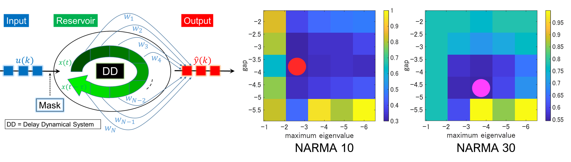
Understanding the life phenomena through non-linear science
In our bodies, cells work together for complex processing and control. Developing and analyzing mathematical and physical theories, including non-linear dynamics, allow us to understand the collective mechanisms of these cells.
- I. Kinoshita, A. Akao, S. Shirasaka, K. Kotani, Y. Jimbo. Analysis of reservoir computing focusing on the spectrum of bistable delayed dynamical systems. Electron Comm Jpn. 102, 15-20, (2019)
- K. Kotani, I. Yamaguchi, Y. Ogawa, Y. Jimbo, H. Nakao, and G. Bard Ermentrout, Adjoint Method Provides Phase Response Functions for Delay-Induced Oscillations, Physical Review Letters Vol. 109, 044101, 2012
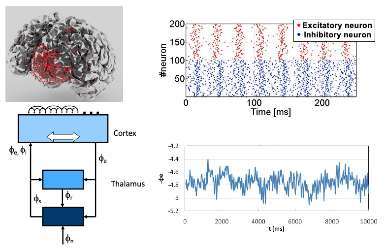
Multiscale mathematical model of the brain
It is a tough challenge to understand the functioning of the brain, which is composed of a vast number of neurons. We are, therefore, constructing a multiscale mathematical model of the brain to illustrate experimental results and/or predict the multiscale dynamics from the level of individual neurons to the level of the entire brain.
- A. Akao, Y. Ogawa, Y. Jimbo, B. Ermentrout and K. Kotani; Relationship between the mechanisms of gamma rhythm generation and the magnitude of the macroscopic phase response function in a population of excitatory and inhibitory modified quadratic integrate-and-fire neurons, Physical review E 97, 012209 (2018)
- Kotani K., Yamaguchi I., Yoshida L., Jimbo Y., Ermentrout G. B., Population dynamics of the modified theta model: macroscopic phase reduction and bifurcation analysis link microscopic neuronal interactions to macroscopic gamma oscillation, Journal of the Royal Society of Interface, Vol. 11, 20140058, 2014
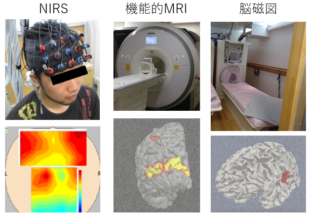
Non-invasive measurement of the human brain
We employ non-invasive measurements of the human brain, including EEG (electro-encephalogram), NIRS (near infrared spectroscopy), and fMRI (functional magnetic resonance imaging). We then seek to illustrate brain functions, such as working memory, calculation, and attention. In addition, we are working on a new methodology to evaluate the human brain non-invasively.
- Ogawa Y., Kotani K., Jimbo Y., Relationship between working memory performance and neural activation measured using near-infrared spectroscopy, Brain and Behavior, Vol. 4, pp. 544-551, 2014
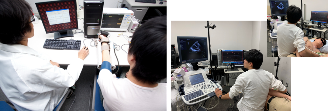
Evaluation of cardiovascular regulation and its application in health care systems
The cardiovascular system constantly delivers blood to the organs in the required amount. Hence, a loss of function may have severe medical consequences, such as heart attacks and cerebrovascular accidents. We propose a robotic system that automatically evaluates vessel elasticity at home to prevent such diseases. We also evaluate how the autonomic nervous system regulates the cardiovascular system, and apply this knowledge in both health care systems and rehabilitation.
- Nishi H*, Mizuno S, Fujino K, Loe, I. A, Wang Y, Ishide T, Jimbo Y, Nangaku M, and Kotani K*, Motion-capture technique-based interface screen displaying real-time probe position and angle in kidney ultrasonography, Clinical and Experimental Nephrology 26, 735-740, 2022
*Equal contribution - Kotani K., Takamasu K., Jimbo Y., Yamamoto Y., Postural-induced phase shift of respiratory sinus arrhythmia and blood pressure variations – insight from respiratory-phase domain analysis. American Journal of Physiology, Heart and Circulatory Physiology, Vol. 294, H1481-H1489, 2008
Enhanced brain-computer interface by multimodal augmented reality techniques
A brain-computer interface is a human support system that decodes the user’s intentions directly from brain signals. In order to increase the reliability of the system with a user-friendly interface, we present stimuli via multiple sensory systems based on augmented reality techniques, and read the user’s intentions from the reaction in his/her brain signals.
- Naito G., Yoshida L., Numata T., Ogawa Y., Kotani K., Jimbo Y., Simultaneous Classification of Multiple Motor Imagery and P300 for Increase in Output Information of Brain-Computer Interface, Electronics and Communications in Japan, Vol. 98, pp. 47-54, 2015
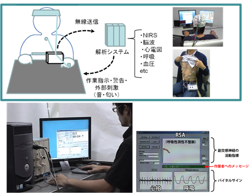
Biofeedback systems for human support
Biofeedback systems are a beneficial support to humans in many aspects of life, such as prevention of stress at work, health care, rehabilitation, and relaxation.
We develop biofeedback systems that are governed by real-time signal processing, and apply them to various fields, including workers’ health care, home relaxation support, and a tool for interactive art in which computer graphics reflect the viewers’ vital signs.
- T. Numata, Y. Ogawa, L. Yoshida, K. Kotani, and Y. Jimbo, High-Accuracy Extraction of Respiratory Sinus Arrhythmia with Statistical Processing Using Normal Distribution, Electronics and Communications in Japan, 96, 23-32, 2013
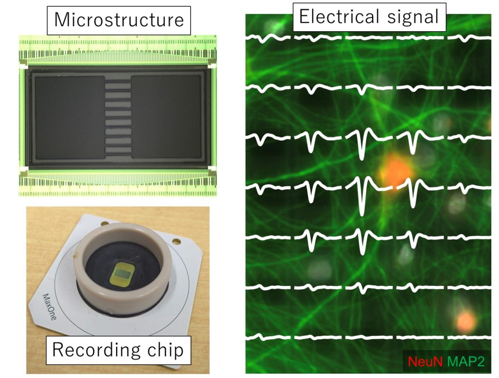
Electrical measurement with high spatiotemporal resolution
The brain functions are expressed with complicated networks formed by a large number of neurons. To understand complex brain functions from a simpler system, we measure the activity of cultured neurons using high density microelectrode array (HD-MEA) chips with 26,400 electrodes. After measurement, targeted proteins are stained to evaluate the relationship between cell type and function.
1) S. Iida, K. Shimba, K. Sakai, K. Kotani, Y. Jimbo, Synchronous firing patterns of induced pluripotent stem cell-derived cortical neurons depend on the network structure consisting of excitatory and inhibitory neurons, Biochemical and Biophysical Research Communications, Vol. 501, Issue 1, pp. 152-157, 2018
2) Y. Tanaka, T. Isomura, K. Shimba, K. Kotani, Y. Jimbo, Neurogenesis enhances response specificity to spatial pattern stimulation in hippocampal cultures, IEEE Transaction on Biomedical Engineering, Vol. 64, Issue 11, pp. 2555-2561, 2017.
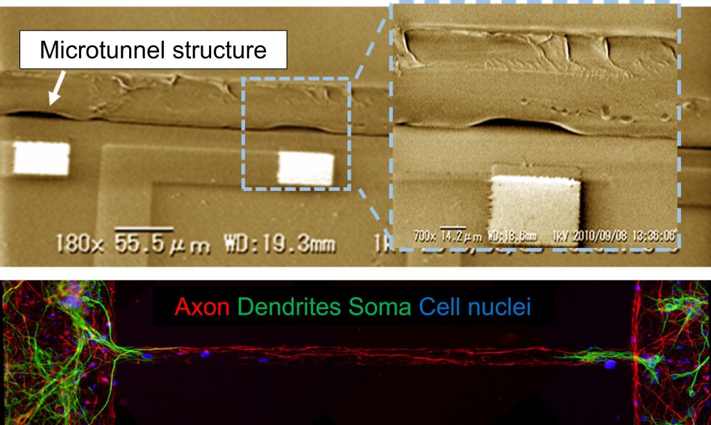
Micro-fabricated device for controlling network structure
Microfabrication technology enables the fabrication of small tunnel structures. By connecting culture compartments with microtunnels, the interaction of multiple networks can be measured. For example, when the hippocampus and cortex networks were connected, each network showed synchronized activity while maintaining its original activity characteristics.
1) C.H. Chang, T. Furukawa, T. Asahina, K. Shimba, K. Kotani, Y. Jimbo, Coupling of in vitro neocortical-hippocampal coculture bursts induces different spike rhythms in individual networks, Frontiers in Neuroscience, Vol. 16, 873664, 2022
2) K. Shimba, K. Sakai, Y. Takayama, K. Kotani, Y. Jimbo, Recording axonal conduction to evaluate the integration of pluripotent cell-derived neurons into a neuronal network, Biomedical Microdevices, Vol. 17, No. 5, 94, 2015
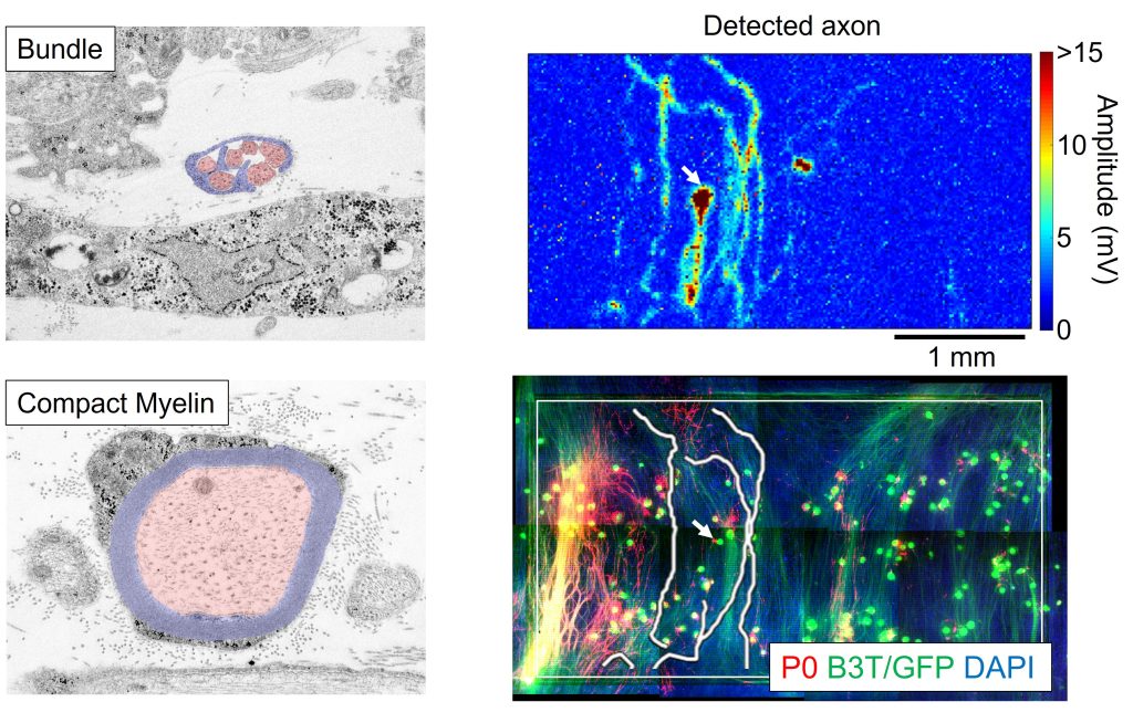
Evaluating axon functions for studying neurological diseases
Taking advantage of the high density of electrodes, we can measure the function of axons, the cables through which neurons send signals to other cells. We have succeeded in reproducing and measuring a phenomenon called “saltatory conduction” in vitro.
1) K. Shimba, T. Asahina, K. Sakai, K. Kotani, Y. Jimbo, Recording Saltatory Conduction along Sensory Axons Using a High-Density Microelectrode Array, Frontiers in Neuroscience, Vol. 16, 854637, 2022
2) K. Shimba, K. Kotani, Y. Jimbo, Microfabricated Device to Record Axonal Conduction under Pharmacological Treatment for Functional Evaluation of Axon Ion Channel, IEEE Transactions on Biomedical Engineering, Vol. 68, No. 12, 2021
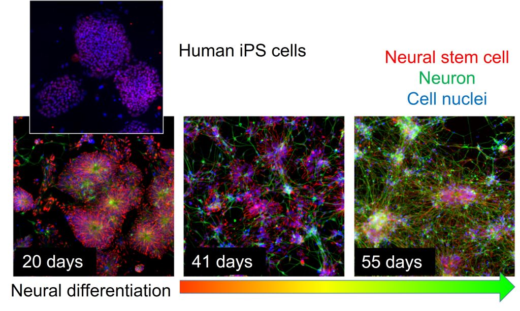
Understanding neural function using human iPS cells
By inducing neural differentiation toward the cerebral cortex, sensory neurons, and spinal cord from human iPS cells, we can obtain cells with human characteristics that are different from those of rats and mice. The application of human iPS cells is to develop human-specific functions and to construct an evaluation system for transplantation of human cells. We have measured that the maturation of human neuronal networks takes about 10 times longer than that of rats.
1) K. Shimba, K. Sakai, S. Iida, K. Kotani, Y. Jimbo, Long-term developmental process of the human cortex revealed in vitro by axon-targeted recording using a microtunnel-augmented microelectrode array, IEEE Transaction on Biomedical Engineering, Vol. 66, No. 9, pp. 2538-2545, 2019
2) S. Iida, K. Shimba, K. Sakai, K. Kotani, Y. Jimbo, Synchronous firing patterns of induced pluripotent stem cell-derived cortical neurons depend on the network structure consisting of excitatory and inhibitory neurons, Biochemical and Biophysical Research Communications, Vol. 501, Issue 1, pp. 152-157, 2018
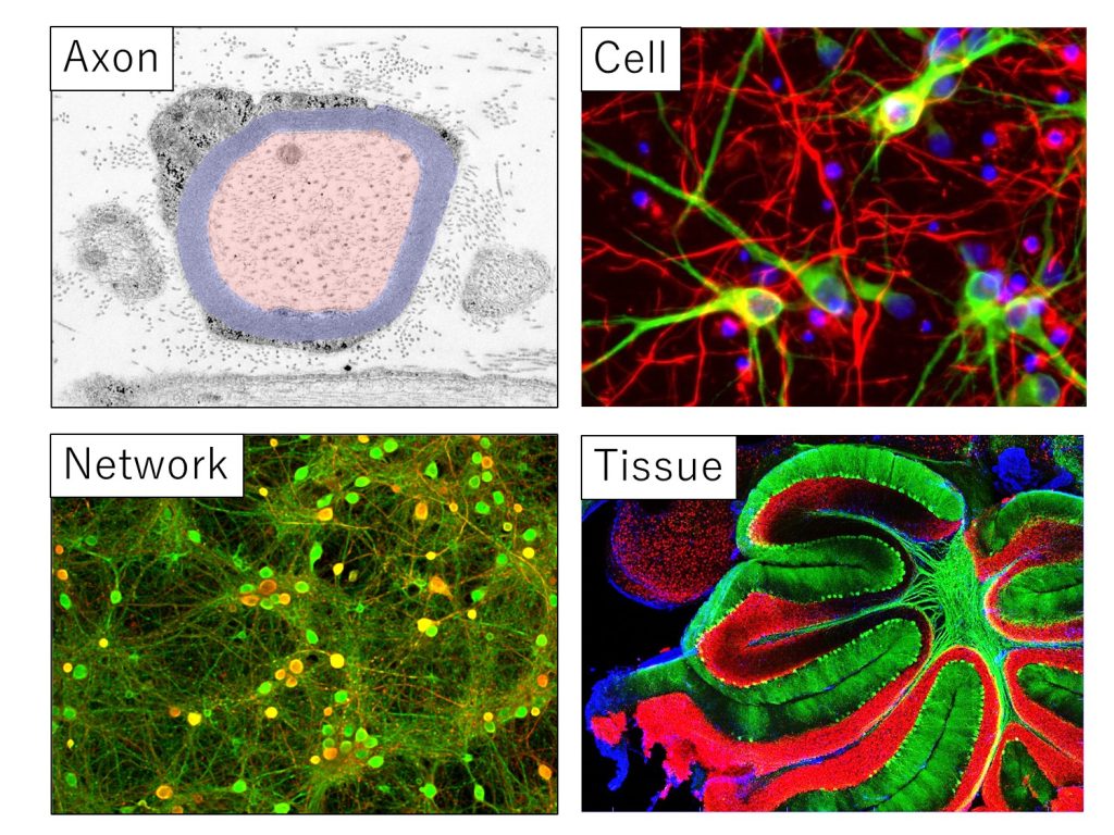
Developing methods for linking neural data on multiple scales
From axons to tissues, we can evaluate function by electrical activity and measure structure by staining from samples of various sizes. Combining these data, we are developing methods for comprehensive measurement and analysis of functions from cells to networks.